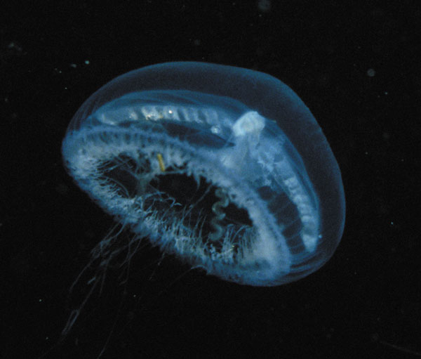
After hearing the music of Calmodulin,I became interested in exploring the musical properties of other calcium-binding proteins. The picture of a jellyfish at the left was taken from a recent article in Nature describing the tertiary structure of one of the Aequorins: (James F. Head*, Satoshi Inouye,Katsunori Teranish and Osamu Shimomura. The crystal structure of the photoprotein aequorin at 2.3 Å resolution. Nature 405 (18 May 2000): 372 - 376.) For more information on Aequorea, Aequorin and its associated protein GFP, see this article by C.E. Mills.
The 3-Dmodel of Aequorin displayed here is from a different member of the Aequorin family. The primary structure of this Aequorin, on which the music is based, is given in the table below, with Calmodulin for comparison. Aequorin feature markings are from files at UniProt and PDB. (Errors of interpretation are, of course, mine.) Since my interest was in the Ca++ binding regions, I have set each binding region on a separate line, even when doing so broke a region of helix. Whereas its sister molecule Calmodulin has four Ca++ binding sites, Aequorin has only three, having apparently lost one since the genes for the two proteins diverged from their common ancestor. Aequorin is also a larger molecule than Calmodulin.
Calmodulin Homo sapiens (human) ( ) = alpha helix | Aequorea victoria (jellyfish) ( ) = alpha helix |
ADQL (TEEQIAEFKEAFSLF) D{KD}GDG[TI]T(TKE LGTVMR) {SLG}QNPT (MKDTDSEEEIREAFRVF) {LG}EKLT(DEEVDEMIREA) | (NPRWIGRHKHMFNFL) D{VN}HN[G]KIS(LDE MVYKASDIVIN) {N}[L]GAT {KN}EPT(LIRIWGDALFDIV) GIIQS(SEDCEETFRVC) |
Notes on the Music:
The music begins with a long introduction in which the theme of the third Ca++-binding domain of the Aequorin sequence is reiterated several times, in several overlapping voices. The entire Aequorin sequence is then introduced on a key change, as the Ca++-binding theme gradually fades. Having heard Ca++-binding site 3 several times, you will recognize its appearance in the full Aequorin sequence. After the full sequence is repeated, it returns, this time with the alpha, beta and turn regions sounding in different voices: the alpha helix regions are clearly marked by a parallel voice set a fourth above the main melodic line played by marimba, and the turns by wood blocks. After this second reiteration of the full aequorin sequence, the sister protein Calmodulin drops in for a brief appearance. Aequorin then returns with fragments of the Ca++-binding theme to finish the piece.
![]()